 how-to
Can Implants Work After Bone Loss? What to Know in 2026
Table of Contents
- How Bone Loss Changes Your Jaw
- Can Implants Work After Bone Loss?
- Bone Grafting for Dental Implants: How It Rebuilds Your Jaw
- Dental Implant Bone Loss Success Rate: What the Research Shows
- Zygomatic Implants for Severe Bone Loss
- The Emotional Side of Bone Loss and Tooth Replacement
- Caring for Grafted Bone and Implants Long Term
- Frequently Asked Questions
Last Updated: September 13, 2026
How Bone Loss Changes Your Jaw
Bone loss after tooth loss is predictable: when a tooth is removed, the alveolar bone that anchored it loses the mechanical stimulation it needs to maintain density, so the jaw gradually resorbs and shrinks.
It accelerates fastest in the first year. The mandibular bone and maxillary ridge lose height and width, changing bite alignment, shifting remaining teeth, and altering facial contours. Long-term denture wearers often reach the point where their dentures no longer fit.
That explains the mechanics. Here is where it gets interesting: the bone that remains is often still usable. Density matters more than total volume, and a cone beam imaging overview from the American Dental Association gives your clinician the three-dimensional picture needed to judge whether implants can work after bone loss.
Can Implants Work After Bone Loss?
Yes. Implants can work after bone loss, but it depends on how much bone remains and where. Implants need enough bone to surround and stabilize the titanium post through osseointegration; when that foundation is insufficient, the jaw can often be rebuilt.
- Mild to moderate loss: Implants usually proceed with standard placement.
- Significant loss in the upper back jaw: A sinus augmentation or sinus lift may be required.
- Severe loss in the lower jaw: Ridge augmentation or, in extreme cases, zygomatic implants.
- Widespread full-arch loss: All-on-4 or full-arch restoration designs can work around limited bone.
The critical step is a CBCT scan, which measures bone volume, height, and density in three dimensions. A panoramic radiograph cannot confirm whether a site has enough depth for a standard implant.
Every implant consultation begins with that imaging, because the difference between a straightforward placement and a grafted site is measured in millimeters.
Bone Grafting for Dental Implants: How It Rebuilds Your Jaw
Bone grafting for dental implants is the surgical placement of donor, synthetic, or patient-derived material into a deficient site to rebuild volume and create a foundation for the implant. The graft acts as a scaffold, and over several months the body replaces it with new bone.
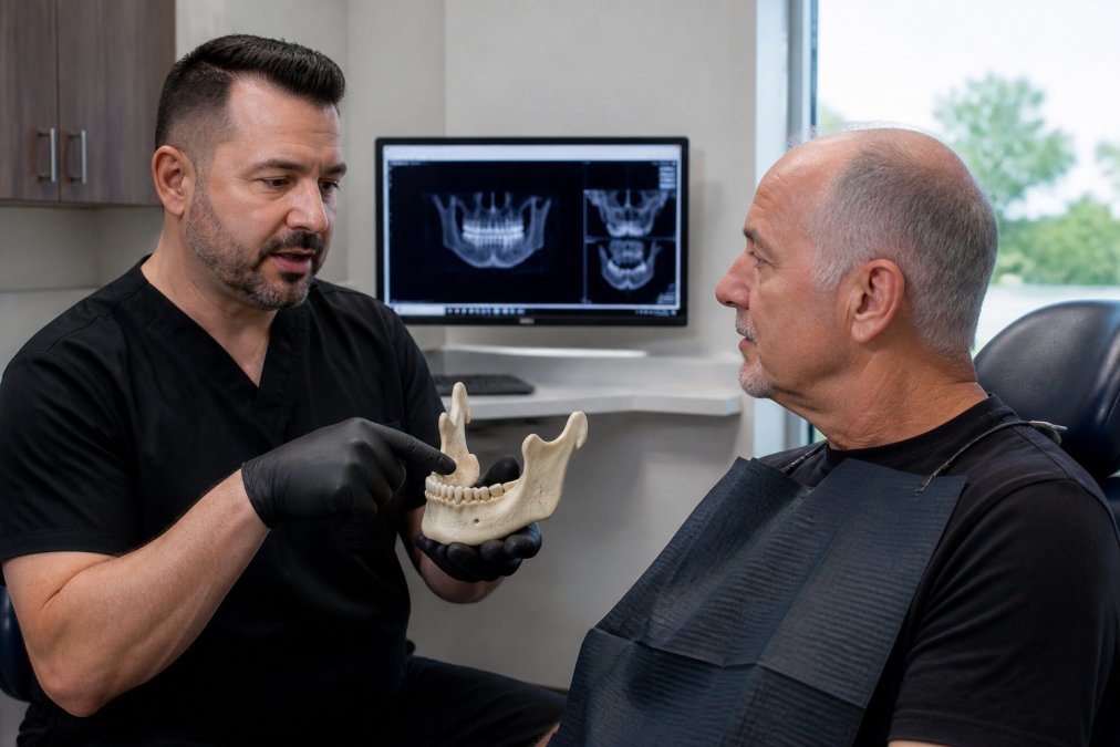
Graft material comes in several forms, chosen by site and the amount of bone needed:
| Graft Type | Source | Common Use |
|---|---|---|
| Autograft | Patient's own bone (often from the chin or ramus) | Larger defects, highest biological activity |
| Allograft | Donated human bone (cadaveric, processed) | Ridge preservation, socket grafting |
| Xenograft | Bovine or porcine bone | Sinus augmentation, ridge augmentation |
| Synthetic bone substitute | Lab-made calcium phosphate or bioactive glass | Smaller defects, socket preservation |
| Composite | Combination of two or more types | Complex or compromised sites |
The Four Procedures Patients Actually Encounter
Bone grafting is an umbrella term. In practice, your clinician will recommend one of four procedures, each with its own timeline and recovery profile.
Socket preservation (ridge preservation) is performed at extraction. Placing graft material into the socket immediately slows resorption and preserves the site for a future implant. It is the lowest-cost, lowest-risk graft because the socket is already open. Ask about it at the extraction appointment, once the ridge has collapsed, a larger augmentation is required.
Ridge augmentation rebuilds width or height in a site that has already resorbed, typically when a tooth has been missing for months or years. The surgeon may use a particulate graft, a block graft, or a membrane to hold the material in place. Healing takes longer than socket grafting because the body must vascularize a larger volume.
Sinus augmentation (sinus lift) addresses the upper back jaw, where the maxillary sinus sits above the molar and premolar roots. After tooth loss, the sinus floor can drop and the ridge resorb from below, leaving too little bone height. A sinus lift elevates the sinus membrane and places graft material beneath it. Two approaches exist:
- Lateral window (external) sinus lift, a small access window is created in the side of the jaw. Used when more height is needed, typically 4 mm or more.
- Transcrestal (internal) sinus lift, the graft is placed through the implant osteotomy site. Used when less height is needed and the remaining bone is adequate to stabilize the implant.
Nerve repositioning or lateralization is a less common lower-jaw procedure used when the inferior alveolar nerve limits available bone height. It is reserved for specific anatomical situations and experienced surgeons.
How Long Grafting Adds to the Timeline
Healing typically takes three to six months before implant placement, varying by graft type, site, and volume. Soft tissue heals within weeks, but the graft must consolidate before bearing an implant:
- Socket preservation at extraction: 3-4 months before implant placement
- Ridge augmentation: 4-6 months
- Sinus lift (lateral): 6-9 months
- Block graft: 4-6 months, sometimes longer
Your surgeon may stage the procedures, graft first, implant later, or place the implant simultaneously when primary stability can be achieved, which shortens the timeline but only works when the remaining bone can hold the implant securely.
What Affects Whether the Graft Takes
Graft success is not automatic. Key factors:
- Blood supply at the site, grafts need vascularity to remodel. Sites with poor blood flow heal more slowly.
- Smoking, nicotine constricts blood vessels and carbon monoxide reduces oxygen delivery, both of which impair graft incorporation.
- Systemic health, uncontrolled diabetes, autoimmune conditions, and certain medications (including some osteoporosis drugs) can affect bone healing.
- Graft material choice, autograft has the highest biological activity but requires a second surgical site; synthetic materials are more predictable in supply but may remodel differently.
- Site stability, a graft that is not protected by a membrane or stable soft tissue can be disrupted during healing.
If you smoke, your surgeon will likely ask you to stop for a defined window before and after surgery, one of the most reliable predictors of graft success.
What Recovery Actually Looks Like
Most patients have swelling, minor discomfort, and tenderness at the graft site for a few days. Swelling peaks at 48-72 hours and subsides over the following week; soft tissue heals in two to three weeks. The graft itself remodels for months, which is why the implant is not placed immediately.
Diet is usually soft and cool for two weeks, advancing as comfort allows. Avoid chewing on the graft site, smoking, and straws (which can dislodge a clot). Follow-up CBCT imaging confirms consolidation before implant placement.
The Lifestyle Factors That Shape Bone Regeneration
The months after surgery are when lifestyle choices most influence whether the graft becomes bone.
- Smoking is the single largest modifiable risk factor. It reduces blood flow, impairs osteoblast function, and is associated with higher rates of graft failure and implant loss.
- Nutrition matters more than most patients expect. Adequate protein, calcium, vitamin D, and vitamin C support the collagen matrix and mineral deposition that bone regeneration depends on. Severe deficiencies are rare, but marginal intake can slow healing.
- Blood sugar control is critical for patients with diabetes. Poorly managed glucose impairs healing and raises infection risk. If your A1C is elevated, your surgeon may coordinate with your primary care provider before proceeding.
- Medications, certain drugs, including bisphosphonates and some biologics, affect bone turnover. Your surgeon needs a complete medication list, including anything prescribed for osteoporosis.
- Alcohol, heavy intake impairs bone formation and should be limited during the healing window.
A patient who stops smoking, manages blood sugar, and eats well during healing gives the graft the best chance of becoming bone that can hold an implant for decades.
Dental Implant Bone Loss Success Rate: What the Research Shows
The dental implant bone loss success rate is high when the site is properly prepared. Implants in grafted bone integrate at rates comparable to native bone, provided the graft has consolidated and periodontal health is stable. Most long-term studies report survival rates above 90% at 10 years, with grafted sites in healthy patients performing similarly.
But "success rate" collapses several questions into one number:
- Survival, is the implant still in the mouth?
- Success, is the implant functioning, stable, and free of peri-implant disease?
- Patient satisfaction, does the restoration look and feel acceptable?
An implant can survive for years while failing on the other two measures. The more useful question is what makes an implant succeed or fail in your situation.
The Factors That Actually Drive Outcomes
Several factors influence whether an implant in grafted bone succeeds:
- Graft maturity: Placing an implant too early into an immature graft increases the risk of failure. The graft must be consolidated enough to provide primary stability.
- Primary stability at placement: This is one of the strongest predictors of long-term success. If the implant can be torqued to an adequate level and shows good initial stability, the odds improve significantly.
- Smoking: Tobacco use constricts blood vessels and slows bone regeneration. Smokers have higher rates of both early and late implant failure.
- Uncontrolled diabetes: Poorly managed blood sugar impairs healing and raises infection risk. Well-controlled diabetes is generally not a contraindication.
- Oral hygiene: Peri-implantitis, the inflammatory condition around implants, is the leading cause of late implant failure. It is largely preventable with consistent maintenance.
- Periodontal history: Patients with a history of severe gum disease are at higher risk of peri-implantitis and need more frequent monitoring.
- Bite forces and parafunction: Heavy clenching or grinding can overload an implant and contribute to bone loss around it.
- Restoration fit: A poorly fitting crown or prosthesis creates a ledge where bacteria accumulate and can lead to crestal bone loss.
A well-maintained implant in a healthy patient with adequate bone behaves very differently from one placed into a compromised site in a patient with untreated gum disease. Most failures trace back to patient selection and maintenance, not to the graft itself.
Early vs. Late Failure: Two Different Problems
Implant failures fall into two categories:
Early failure occurs before the implant has fully integrated, usually within the first few months. The most common causes are inadequate primary stability, infection, premature loading, and placement into a graft that had not consolidated. Early failure is often a surgical or timing issue.
Late failure occurs after the implant has integrated and is in function, sometimes years later. The dominant cause is peri-implantitis, inflammation around the implant that leads to progressive bone loss, plus overload from bite forces, poor restoration fit, and untreated systemic conditions. Late failure is largely a maintenance issue. Prevention differs: early failure is reduced by careful surgical planning and imaging; late failure by consistent hygiene, professional maintenance, and prompt treatment of inflammation.
What the Timeline Looks Like
Osseointegration, bone growing into and around the implant surface, typically takes three to six months in the lower jaw and four to six in the upper. Grafted sites may take longer because the graft must consolidate first, and loading too early is a known cause of failure. After restoration, the bone around the implant responds to load, helping maintain density as natural teeth do. The vulnerable window is between extraction and implant placement, when the unloaded site resorbs faster.
Long-Term Maintenance: What Keeps Grafted Bone and Implants Healthy
Long-term maintenance determines whether the investment holds. Grafted bone and implants need the same care as natural teeth, plus a few additions:
- Professional cleanings every six months, or more often if you have a history of periodontal disease or peri-implantitis.
- Daily interdental cleaning with brushes or floss designed for implant restorations. Standard floss can be awkward around implant crowns; interdental brushes and water flossers are often more effective.
- Regular imaging to monitor bone levels around the implant and detect peri-implantitis early. Early bone loss is often asymptomatic, which is why imaging matters even when nothing feels wrong.
- Lifestyle adjustments that support bone health: not smoking, managing blood sugar, and adequate calcium and vitamin D intake.
- Bite monitoring, if you clench or grind, a night guard may be recommended to protect the implant and restoration from overload.
Lifestyle factors affecting bone regeneration are real and modifiable. Smoking is the biggest controllable risk factor; nutrition and blood sugar control matter more than most patients expect. Grafted bone also responds to load: once an implant is functioning, the surrounding bone is stimulated again, helping maintain density. The years between extraction and placement are the vulnerable window; the years after restoration are when maintenance habits determine how long the result lasts.
When to Get a Second Opinion
If you have been told you are not a candidate because of bone loss, or have an implant showing trouble, a second opinion with three-dimensional imaging is reasonable. CBCT reveals bone volume and density a panoramic radiograph cannot, and many patients told they had "no bone" turn out to have enough for a grafted or angled implant. The goal is not to shop for a yes, it is to base the decision on complete diagnostics.
Zygomatic Implants for Severe Bone Loss
Zygomatic implants for severe bone loss anchor into the zygomatic bone (cheekbone) rather than the upper jaw, bypassing sinus augmentation when maxillary bone is too resorbed for conventional implants.
The zygomatic bone is dense and rarely resorbs, even after decades of edentulism, making it a reliable anchor when the upper jaw has atrophied. The tradeoff is a more complex procedure with a longer learning curve, and not every practice offers it.
For full-arch upper restorations with severe atrophy, zygomatic implants can mean fewer surgeries and, in some protocols, same-day teeth. The alternative is often staged: sinus lift, graft healing, then implant placement, stretching the timeline considerably. Candidacy requires careful imaging and a clinician experienced with the technique, this is not a procedure to select based on convenience alone.
The Emotional Side of Bone Loss and Tooth Replacement
Bone loss carries a psychological weight most clinical discussions skip. Patients describe avoiding photographs, hiding their smile, and feeling older than they are; because the loss is gradual, the toll accumulates quietly. The anxiety often centers on being judged: patients who delayed treatment for years worry a dentist will lecture them or that the damage is too far gone, and that fear keeps people away from consultations that could help. Addressing it starts with a conversation, not a procedure, understanding the timeline, the steps, and what is realistically achievable removes much of the dread. If anxiety has kept you out of the chair, say so at the first visit; a practice equipped to handle it will slow down and explain.
Caring for Grafted Bone and Implants Long Term
Bone loss after tooth loss is common, but it does not close the door on implants. The path forward depends on accurate imaging, the right graft or implant design for your anatomy, and a maintenance plan you can keep. The path forward depends on accurate imaging, the right graft or implant design for your anatomy, and a maintenance plan you can keep. If you have been told you are not a candidate, get a second opinion with proper three-dimensional imaging before you accept that answer.
Frequently Asked Questions
Is it possible to get dental implants if I have bone loss?
Yes, in many cases. Bone loss after tooth loss is common, but it does not automatically rule out implants. A surgeon can rebuild jaw volume with bone grafting for dental implants, or use longer implants anchored in the zygomatic bone when the upper jaw has severe resorption. A CBCT scan and panoramic radiograph measure your jawbone density and bone volume to confirm candidate eligibility. Some patients qualify for standard implants after grafting; others need zygomatic or full-arch solutions. Only an in-person exam can tell you which path fits your anatomy.
What is a bone graft for dental implants?
A bone graft adds material to your jaw so there is enough bone volume for an implant to fuse with. Graft types include autograft (your own bone), allograft (donated human bone), xenograft (bovine or porcine), and synthetic bone substitute. The graft supports new bone growth through a process called bone regeneration. Ridge preservation is a related procedure done right after a tooth is removed to limit alveolar bone loss. Healing usually takes several months before implant placement, depending on the graft size and your periodontal health.
How much bone is needed for a dental implant?
There is no single number that applies to everyone. Your surgeon evaluates jawbone density, bone volume, and the position of the maxillary sinus or mandibular nerve using a CBCT scan. As a general rule, the implant needs enough bone to surround and stabilize it without touching nerves or sinuses. If you are short on bone, a graft, sinus augmentation, or a zygomatic implant can bridge the gap. Implant stability at placement is the practical test your surgeon uses to decide whether to load the implant the same day or wait.
How long does it take for bone to heal after a graft?
Most grafts need three to six months before an implant can be placed, though larger grafts or sinus augmentation may take longer. Soft tissue healing happens in the first few weeks; the grafted bone continues to consolidate for months after that. Lifestyle factors affect bone regeneration, so avoid smoking, keep blood sugar controlled if you have diabetes, and follow your surgeon's instructions on diet and hygiene. Your surgeon will confirm readiness with imaging before the next stage. Rushing the timeline can compromise osseointegration.